How to Use Humeral Interlocking Nail for Bone Fractures?
Bone fractures are common injuries that require effective treatment for proper healing. Among the various methods, the use of a Humeral Interlocking Nail stands out. This device is designed to stabilize the humerus bone during recovery, ensuring alignment and support.
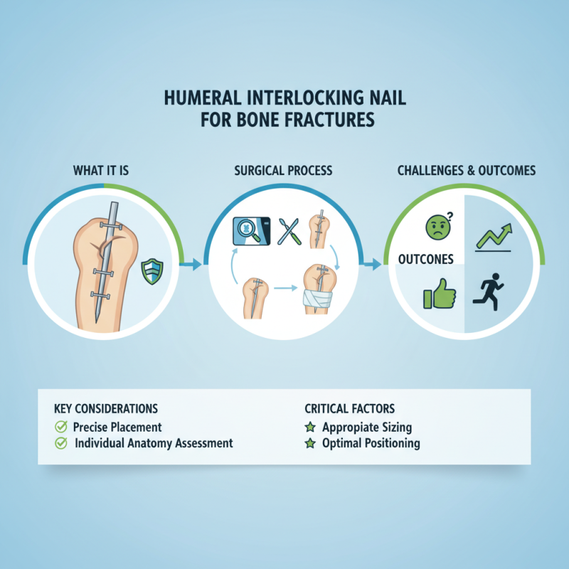
When using a Humeral Interlocking Nail, careful consideration is essential. The surgical process involves precise placement to avoid complications. Surgeons must assess each case individually, as anatomy can vary significantly. Appropriate sizing and positioning of the nail are critical.
Despite its advantages, the method is not without challenges. Surgeons may face difficulties during the insertion process. Understanding the unique characteristics of each fracture is vital. Reflecting on past experiences reveals that not every case is straightforward. The learning curve may be steep, but mastering this technique can lead to better patient outcomes.
Understanding Humeral Interlocking Nails and Their Applications
Humeral interlocking nails are crucial in treating bone fractures. These implants help stabilize the humerus, promoting healing. They are specially designed to interlock with screws, providing strong support. Surgeons often use these nails for complex fractures. They allow for early mobility and reduce complications.
When considering surgical options, it’s vital to understand how the nails function. They are inserted into the bone’s medullary canal. This allows for a minimally invasive approach. However, improper placement can lead to complications. Surgeons must take care to assess the fracture type thoroughly.
Tips: Always consult with your medical team about risks. Recovery differs for everyone. Be honest about your rehabilitation progress. Your feedback can enhance the treatment plan. Regular follow-ups are essential to check the healing. Engaging in physical therapy can be beneficial. It aids the recovery process, making mobility easier.
Preparing for Surgery: Patient Evaluation and Imaging Techniques
Preparing for surgery is a crucial step in treating bone fractures with a humeral interlocking nail. Patient evaluation begins with a comprehensive medical history. Surgeons must assess underlying health conditions that might influence recovery. This includes any history of osteoporosis, diabetes, or autoimmune disorders. Research indicates that fractures occur in nearly 18% of the adult population each year. Understanding the patient's overall well-being can enhance surgical outcomes.
Imaging techniques are vital for accurate diagnosis. X-rays are standard for visualizing bone alignment and fracture severity. However, computed tomography (CT) scans provide more detail. They can identify complex fractures or assess soft tissue involvement. A study found that CT reduces the risk of postoperative complications by 25%. Radiologists play a key role in interpreting these images, ensuring surgeons have clear insights before the procedure.
Tips for evaluation include asking about the patient's pain level and mobility. This helps gauge fracture impact on their daily life. Educate the patient on potential risks of the surgery. Clear communication fosters trust and promotes a smoother surgical experience. Always remember, the patient's comfort and comprehension should be prioritized. Reflecting on how to improve this evaluation process can lead to better-informed decisions in their care.
Surgical Procedure: Step-by-Step Guide to Inserting the Nail
The surgical procedure for inserting a humeral interlocking nail begins with precise planning. Surgeons need to assess the fracture type and location. Imaging studies are essential to visualize the bone structure. Mark the entry point on the humerus, ensuring it's in line with the medullary canal.
Next, a skin incision is made to expose the area. The nail's entry point is reamed carefully to accommodate the implant. During this step, precision is crucial. Mistakes can lead to inaccurate placement. The nail is then inserted through the reamed canal. It should be pushed through until it fits snugly within the bone.
Locking the nail is the final step. Surgeons use screws to secure the nail in position. This prevents any unwanted movement. It's vital to check alignment and stability. Post-operative care includes monitoring for potential complications. Adjustments may be necessary, emphasizing the importance of reflection on each procedure performed.
Post-Operative Care and Rehabilitation for Bone Fractures
Post-operative care for bone fractures treated with a humeral interlocking nail is crucial. After surgery, patients often experience swelling and discomfort. Elevating the arm can help reduce swelling. Ice packs may also be beneficial. Encourage gentle movements, but avoid strenuous activities initially.
Rehabilitation plays a key role in recovery. Physical therapy sessions should start as soon as possible. Patients may feel anxious or frustrated during this process. It’s essential to set realistic goals for mobility and strength. Regular assessment by a physical therapist can guide progress. Monitor pain levels closely and communicate any concerns with healthcare providers.
Patience and persistence are vital during recovery. Each person's healing journey is unique. Celebrate small victories, like increasing range of motion. Reflecting on the rehabilitation experience can help adjust future goals. Gradually incorporating daily tasks into routine can also boost confidence. Always listen to your body and don’t rush the process.
Potential Complications and How to Manage Them During Recovery
Humeral interlocking nails are crucial in treating bone fractures. These devices stabilize the bone during healing. However, complications can arise. One common issue is infection at the surgical site. Reports indicate that infection rates can be as high as 6% in some cases. Proper sterile techniques and follow-up care are essential to minimize this risk.
Another complication is malunion or nonunion of the bone. Research shows that 10-20% of fracture patients may experience this issue. Factors like patient age and nutritional status play significant roles. Encouraging good nutrition and regular check-ups can help monitor healing. Pain management is also critical. Patients may experience severe pain post-surgery, requiring tailored approaches for relief.
Physical therapy is vital during recovery. It aids mobility and strengthens the surrounding muscles. However, some patients may be resistant to following through with therapy, leading to longer recovery times. Open communication between patients and healthcare providers can address concerns and enhance compliance. Overall, understanding these potential complications can lead to better outcomes and smoother recoveries.